


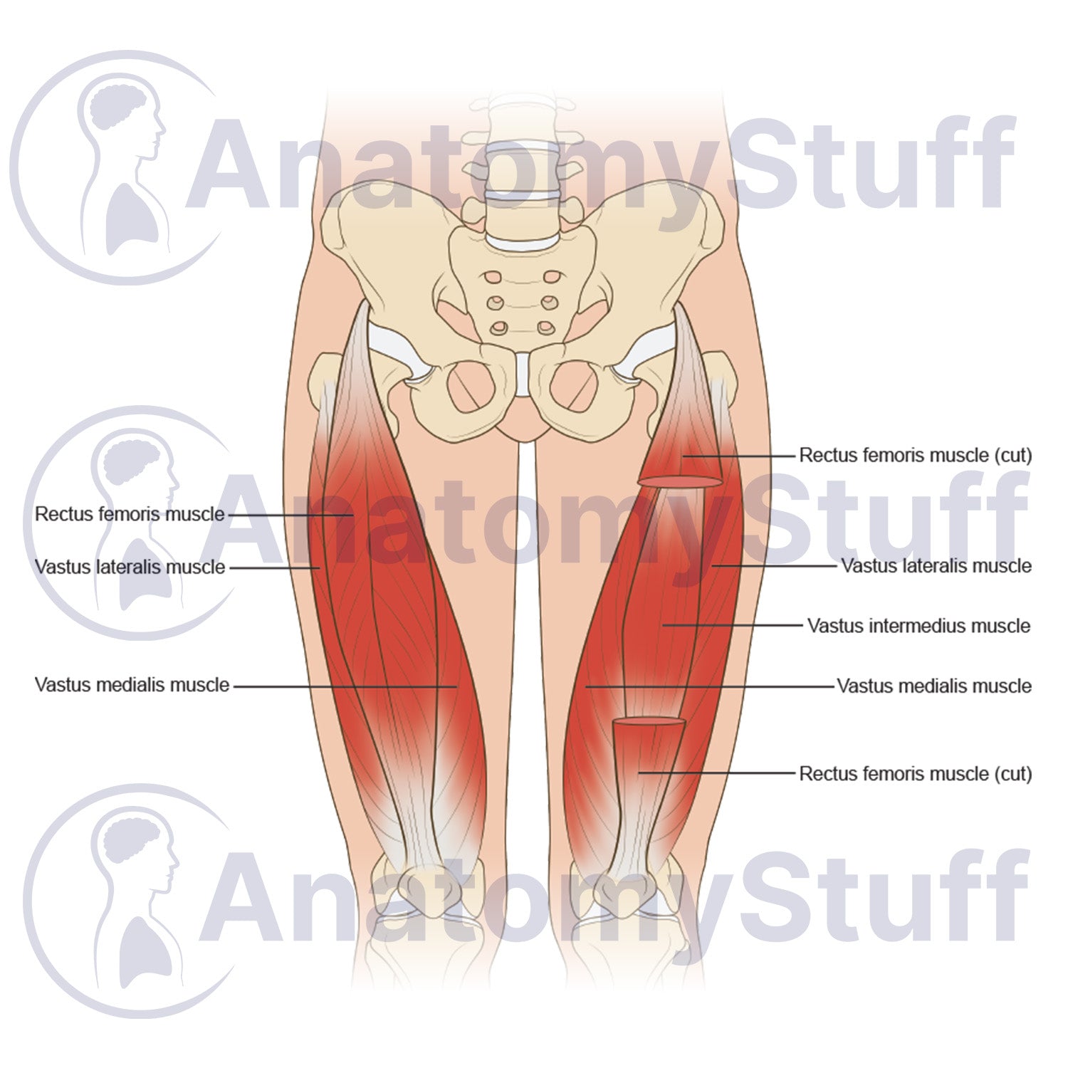
An anatomical quadriceps anatomy stock image detailing the muscle layers and deep structural orientation of the anterior thigh. This professional illustration is designed for university level musculoskeletal study, orthopaedic research publishing, and academic lectures. The layout balances deep structural accuracy with a clear perspective of lower extremity muscle mechanics.
Musculoskeletal Features
- Quadriceps femoris group: Comprehensive diagram tracking the rectus femoris, vastus lateralis, and vastus medialis within the anterior femoral compartment.
- Deep tissue exposure: Specialised dissection view showing the isolated vastus intermedius muscle beneath the sectioned rectus femoris tendon.
- Pelvic alignment: Structural reference mapping muscle pathways against the anterior inferior iliac spine and femoral architecture.
Product Specifications
- Format: PNG (Transparent background)
- Dimensions: 1100 x 800 px
- Resolution: 300 DPI
- Print Size: ~10 x 8 cm
- Colour Profile: RGB (Optimised for digital and print)
- File Size: ~300 KB
Licensing Information
Please select the licence that matches your intended use:
- Science Licence: Licence for academic purposes such as theses research publishing, and the scientific discourse.
- Education Licence: Licence for educational purposes, live teaching, presentations, handouts, and exam papers.
Commercial Use: Interested in using this for advertising, book publication, or other commercial purposes? Please Contact Us to discuss a Commercial Licence.
Please allow 1-2 working days for delivery of your image.
By purchasing, you agree to our Licensing Terms and Conditions.