


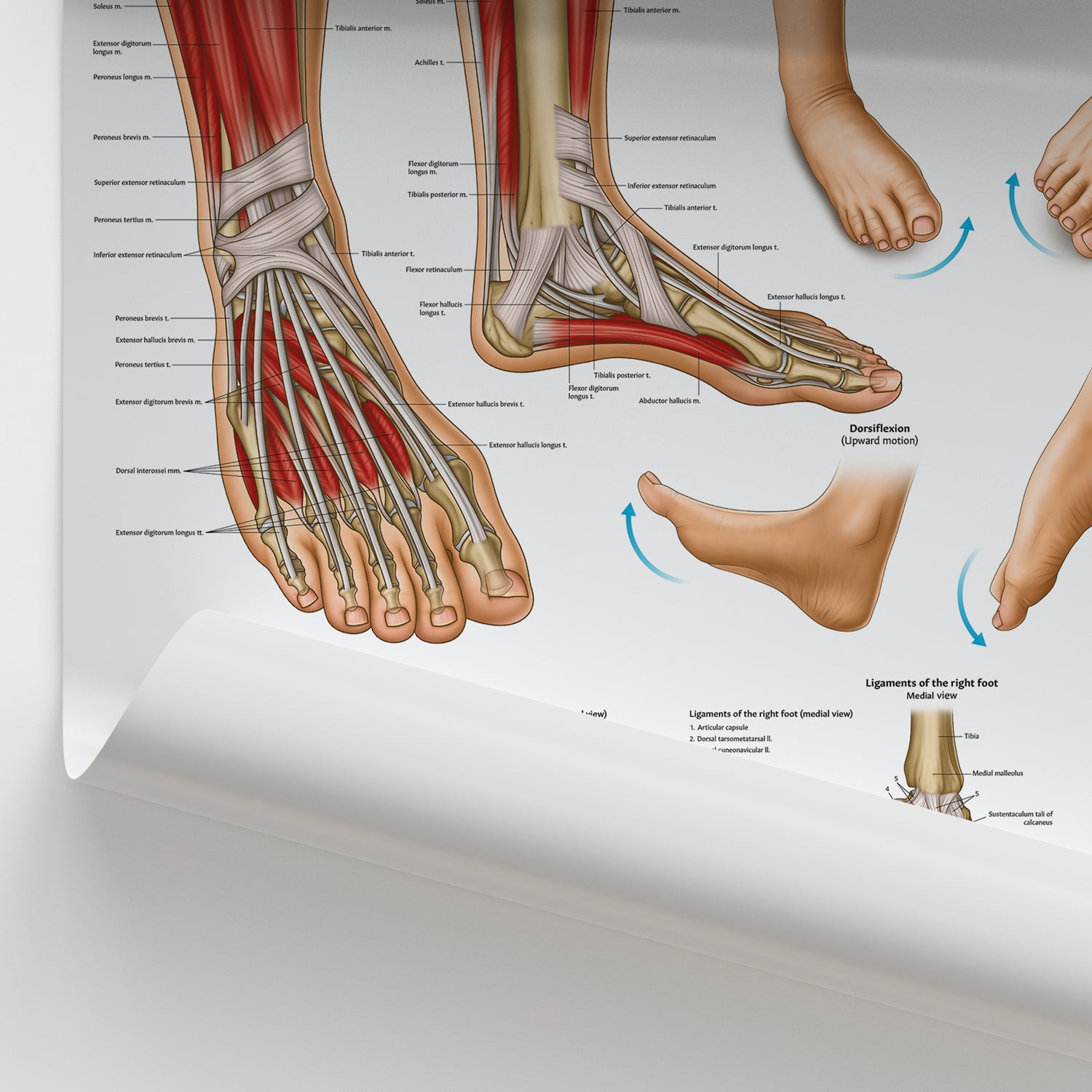


This striking Foot and Ankle Anatomy Chart offers detailed visual insights into the complex structures of the lower limb. Designed for educational and professional use, it's perfect for physiotherapists, sports health practitioners, and students studying anatomy at university level.
Exclusively illustrated by our in-house medical artists and only available at AnatomyStuff.com, this foot and ankle anatomy chart includes:
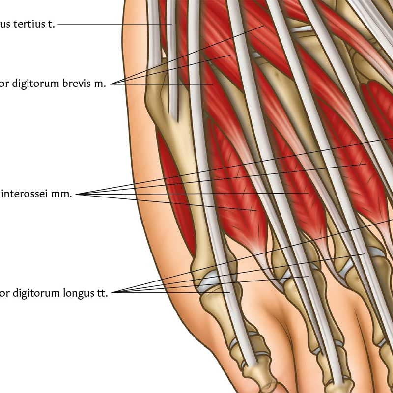
- Lateral and frontal illustrations highlighting muscles, bones, and ligaments
- Dorsal foot view showing skeletal features
- Plantar view focusing on muscular structures
- Comparative diagrams of high and low foot arches
- Common foot movements such as dorsiflexion, plantar flexion, inversion, and eversion
- Ligament structures of the ankle and foot, presented from both medial and lateral perspectives
Our display options suit a variety of clinical and educational settings:
- Classic Semi-Gloss Prints: Offered in 45 x 60 cm, 60 x 80 cm, and 70 x 100 cm sizes, the vibrant semi-gloss finish enhances clarity and colour.
- Framed Prints: Choose from black or white frames, with sizes including 45 x 60 cm, 60 x 80 cm, and 70 x 100 cm. Printed on premium 170 gsm matte paper with a smooth, non-reflective finish and ready to hang in sturdy pine frames.
Add this beautifully rendered Foot and Ankle Joint Anatomy Chart to your clinic or study space—available exclusively from AnatomyStuff.com.