


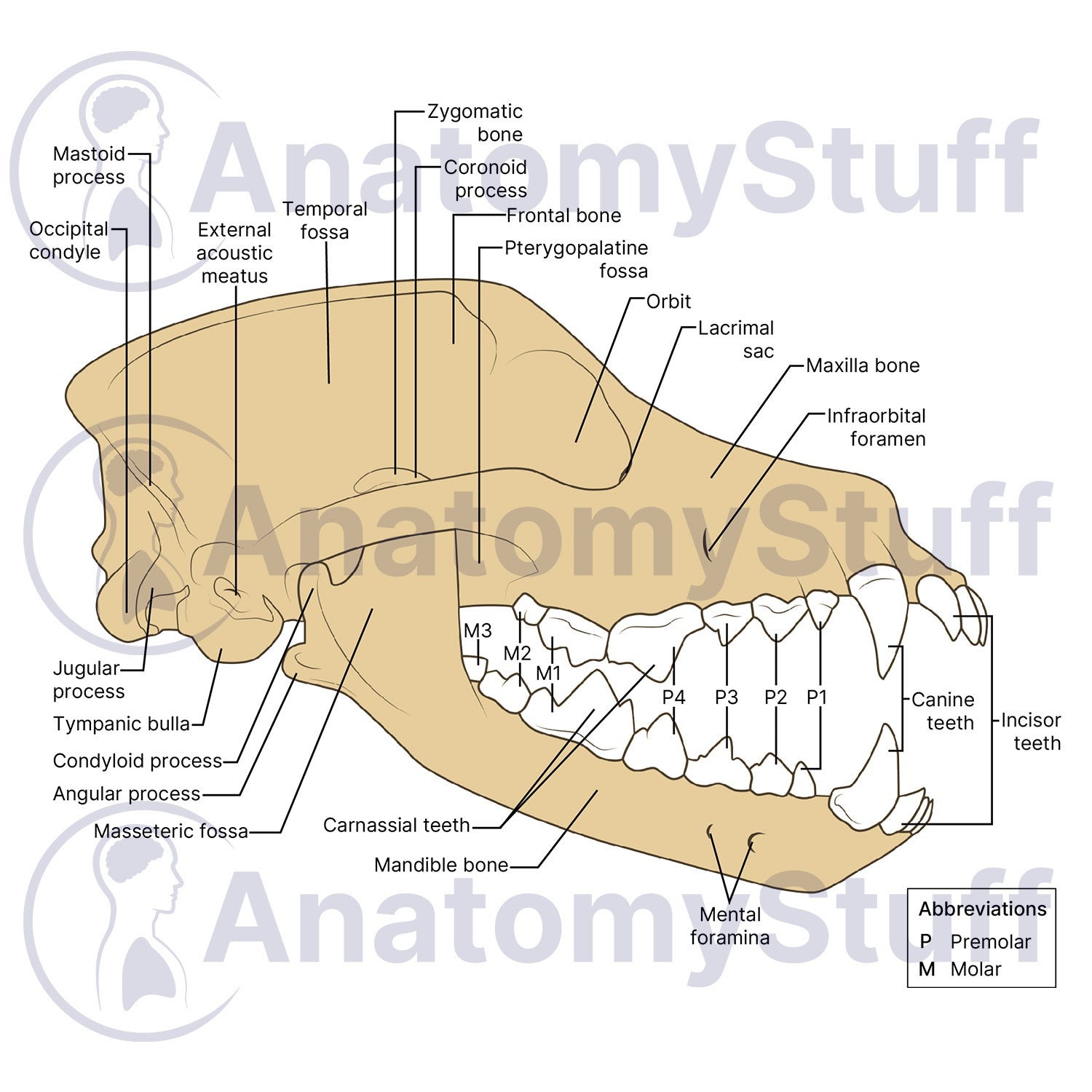
This professional illustration of the Canine Skull Anatomy offers an accurate and detailed anatomical breakdown of a dog skull, ideal for global life sciences, zoology, and veterinary research.
Key Diagram Features & Anatomical Labels
- Cranial & Jaw Architecture: Clear visual mapping of the main skull bones including the frontal, maxilla, and mandible (jaw bone).
- Anatomical Processes & Fossae: Detailed renderings of the temporal fossa, masseteric fossa, tympanic bulla, and the coronoid, condyloid, and angular processes for comparative anatomy studies.
- Foramina & Openings: Highly detailed breakdown of crucial skull passages, clearly marking the infraorbital foramen, mental foramina, and external acoustic meatus.
- Specialized Dentition: Sharp, distinct labeling of canine tooth structure, isolating incisors, canine teeth, premolars, molars, and the specialized carnassial teeth.
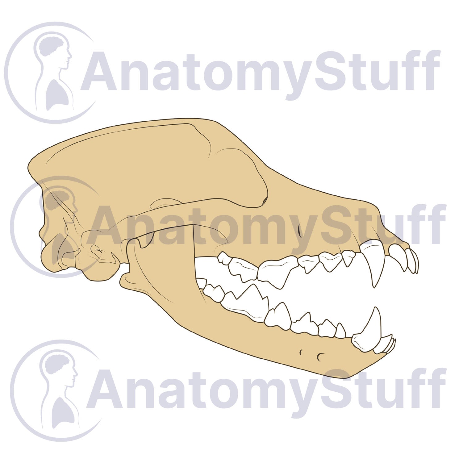
Choose Your Variant
- Fully Labelled: Ready for immediate use in presentations.
- Unlabelled (Blank): Perfect for interactive learning. This clean version is ideal for student examinations, "fill-in-the-blank" quizzes, or custom labelling for specialised research.
Product Specifications
- Format: PNG (Transparent background)
- Dimensions: 1181 x 945 px
- Resolution: 300 DPI
- Print Size: ~ 10 x 8 cm
- Colour Profile: RGB (Optimised for digital and print)
- File Size: ~200 KB (Blank) ~300 KB (Labelled)
Licensing Information
Please select the license that matches your intended use:
- Science Licence: License for academic purposes such as theses research publishing, and the scientific discourse.
- Education Licence: License for educational purposes, live teaching, presentations, handouts, and exam papers.
Commercial Use: Interested in using this for advertising, book publication, or other commercial purposes? Please Contact Us to discuss a Commercial License.
Please allow 1-2 working days for delivery of your image.
By purchasing, you agree to our Licencing Terms and Conditions.